HU
HU
 EN
EN
Musculoskeletal problems
If you experience pain in any of the areas below, feel free to turn to our specialists.
If you experience pain in any of the areas below, feel free to turn to our specialists.
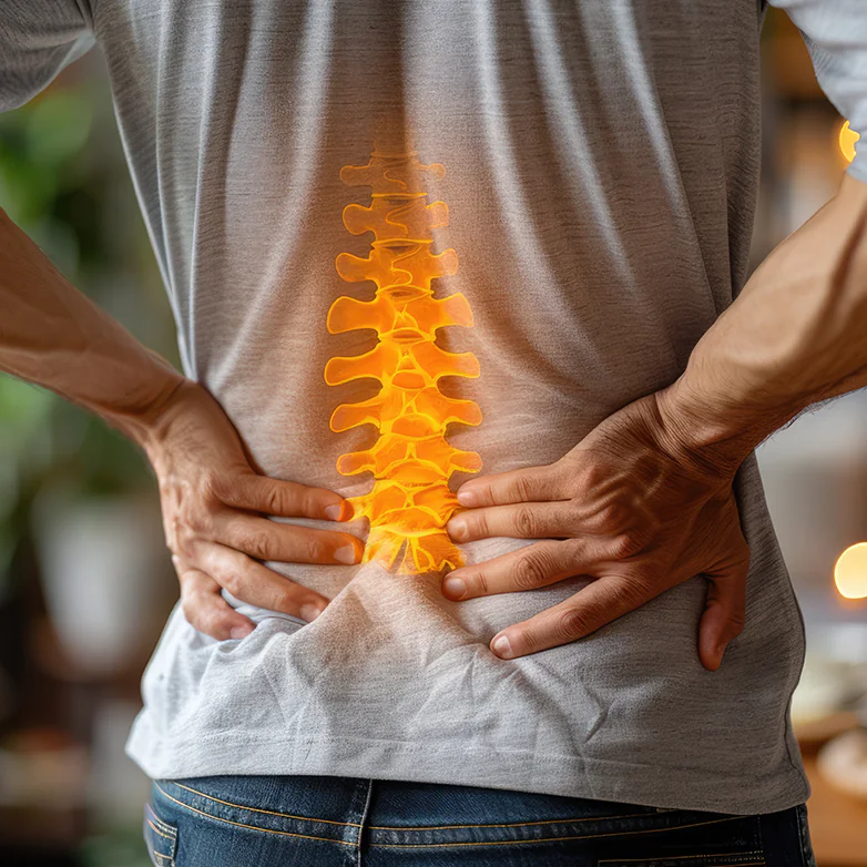
The spine is the body’s main supporting structure, providing stability, protecting the spinal cord, and enabling mobility. It is made up of vertebral bodies separated by intervertebral discs. The vertebrae are connected by small facet joints, and an anterior and posterior ligament system maintains overall integrity. The discs play a key role in allowing flexible movement and absorbing shock. As discs are composed of a jelly-like nucleus surrounded by a fibrous collagen layer, they change shape in response to load — primarily to the pressure generated by gravity. Such changes are frequently described in imaging reports, for example as bulging discs.
The spine is divided into four main sections:
The most common spinal conditions:
The upper limb connects to the trunk via the shoulder joint. The shoulder is a complex and highly mobile joint, allowing the arm to move in almost every direction. However, this very mobility also makes the shoulder vulnerable to injury and inflammation.
Shoulder pain is one of the most common musculoskeletal complaints and can occur at any age. As the pain worsens, shoulder movement gradually becomes restricted, causing significant discomfort during everyday activities such as dressing, grooming, or lifting.
The most common shoulder conditions:
The lower limb connects to the trunk through the hip joint, which plays a key role in movement and maintaining posture. The hip is a ball-and-socket joint formed by the femoral head and the acetabulum of the pelvis, allowing movement in multiple directions: flexion, extension, abduction, adduction, and rotation. It enables walking, running, standing up from a seated position, and many everyday movements.
As the hip bears most of the body’s weight, it is no surprise that hip pain is one of the most common musculoskeletal complaints.
The most common hip conditions:
The health of the hip joint is essential for maintaining mobility and quality of life.
If hip disease progresses to the point where orthopaedic surgery is required, a course of balneotherapy treatments can also contribute effectively to the success of the procedure.
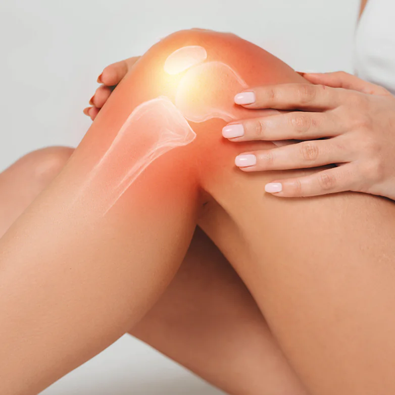
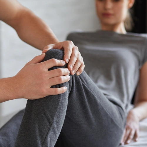
The knee is one of the body’s largest and most complex weight-bearing joints, playing a key role in walking, squatting, climbing stairs, and all movements that require bending or extending the leg. During normal walking, the load on the knee is determined by body weight and the forces acting on the joint, meaning that with each step the knee typically bears around 1.5 times the body weight (for a person weighing 70 kg, this is roughly 105 kg). With faster walking or stair climbing, the load increases to 3–5 times the body weight, and during running it may reach 5–6 times the body weight. As the knee is constantly exposed to load, it is particularly vulnerable to degenerative and inflammatory conditions as well as injuries.
The most common knee conditions:
Bursitis around the knee: inflammation of the bursae, causing swelling and pain.
If knee disease progresses to the point where orthopaedic surgery is required, a course of complex balneotherapy can significantly support the success of the procedure.
During herniated disc surgery, the intervertebral disc material located between the vertebrae is removed. A protruding disc can compress the nerve root, causing severe pain, sensory loss, or difficulty moving the toes or feet (in the case of the lumbar spine). Once the jelly-like disc material is removed during surgery, the pressure on the nerve fibre is relieved, and symptoms usually disappear. However, due to scar tissue formation, additional treatment is often required to restore optimal spinal mobility.
Joint replacement is usually preceded by many years of illness, during which the articular cartilage becomes so worn that the bone ends rub directly against each other during movement. Our vasodilating and circulation-enhancing treatments work exceptionally well on the contracted joint capsule, ligaments, and shortened muscles. They provide excellent surgical preparation and increase the success of the procedure.
Recommended pre-operative programme for joint replacement: thermal water pool therapy, mud treatment, individual underwater physiotherapy, ultrasound therapy.
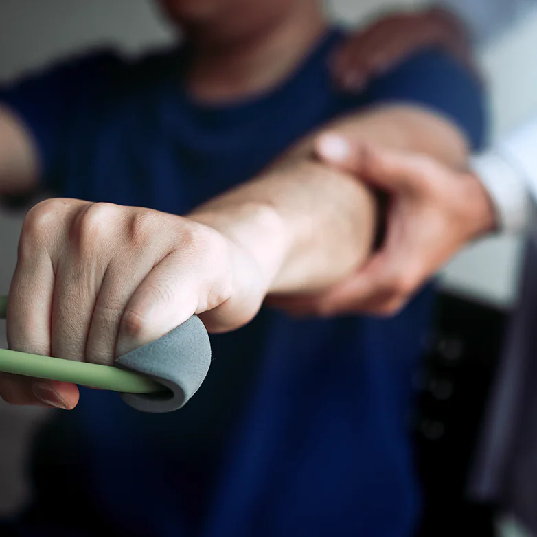
The most common orthopaedic surgeries involve the hip, knee, and shoulder joints. After surgery, the path to full recovery is still long. Early, professional rehabilitation is essential to ensure that the joint regains proper function and the patient can return to their everyday activities.
During early rehabilitation following joint replacement, within the first 6 weeks we combine treatments that do not require aquatic therapy but significantly support recovery.
Recommended dry therapeutic rehabilitation in the first 6 weeks after surgery: individual dry physiotherapy, medical therapeutic massage, soft laser therapy, Bemer vascular therapy.
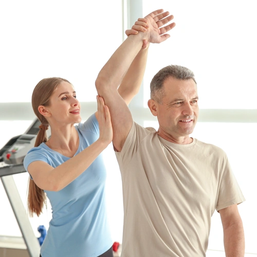
Once the surgical scar has fully healed, rehabilitation should be continued with the combination of thermal water therapy, other hydrotherapeutic procedures, and individual dry physiotherapy in order to regain the best possible range of motion.
Recommended thermal water rehabilitation after the first 6 weeks: thermal water pool therapy, individual underwater physiotherapy, mud pack treatment, individual dry physiotherapy.
Trauma-related surgeries vary widely, but recovery after ankle, wrist, shoulder, and femoral neck fractures represents the majority of cases.

We recommend the services of our spa to anyone
who wishes to maintain or restore their musculoskeletal health — whether to relieve existing complaints or for prevention — regardless of age or life situation.
We welcome those who work in sedentary jobs and often feel tension in their neck, back, or lower back; people who exercise regularly and value proper muscle recovery; individuals doing physical work who place increased strain on their bodies; as well as those in stressful, mentally demanding professions who seek rest and rejuvenation.

The spa is also an ideal choice
if you currently have no complaints but value prevention and wish to maintain your wellbeing in the long term.
Our services can also be an excellent complement for those who have previously received musculoskeletal therapy in a specialist clinic and now wish to maintain or improve their wellbeing in a calm, more natural environment.

If the Széchenyi Medical Spa has sparked your interest, we will help you navigate the various therapies. With an appointment for a specialist examination, we ensure that you receive care without waiting time. A referral is not required for our specialist clinic.

After booking an appointment, you will be seen by one of our rheumatologists or musculoskeletal rehabilitation specialists. The examination includes a physical assessment, which allows us to evaluate joint function and differentiate the sources of pain. For the specialist visit, please bring any previous medical reports related to musculoskeletal or other chronic conditions, so the examining doctor can gain a comprehensive understanding of your health status.

During the specialist examination, you and the doctor will discuss the findings, the nature of your musculoskeletal condition, and the possible treatment options. The specialist will create a therapeutic plan tailored to your specific needs and health status.
Based on the specialist’s therapeutic plan, a personalised treatment schedule will be created — including only those therapies that best suit your condition, goals, and your available time and financial resources.
Treatments can be used independently as non-aquatic therapies — such as medical therapeutic massage, ultrasound, or physiotherapy — but our thermal-water-based therapies are also available. The most effective treatment programmes combine these approaches, creating integrated therapies that act through multiple forms of stimulation.

Throughout the course of treatment, we closely monitor any changes in your condition, and at the end of the programme a specialist follow-up examination is carried out to reassess the results achieved.

Our strategic partner: DOKTOR24
With personalised therapeutic packages created by rheumatologists, orthopaedic specialists, and rehabilitation professionals, we provide comprehensive care.